Introduction
For numerous a long time, electron microscopy has been at the bleeding edge of logical investigate since it offers a never-before-seen see into the nuclear and atomic structure of materials. We’ll look at the most up to date strategies and advancements in electron microscopy in this web journal post, which are progressing science and innovation.
The Evolution of Electron Microscopy
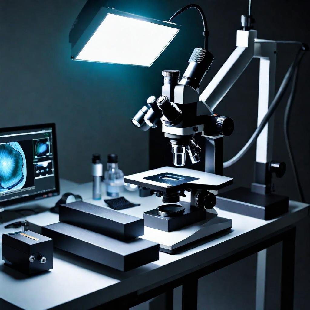
Since its innovation within the early 1900s, the electron magnifying instrument has created into an fundamental instrument in numerous areas, such as materials science, science, and nanotechnology. Since electrons have distant shorter wavelengths than unmistakable light, electron magnifying instruments, in differentiate to customary light magnifying lens, enlighten the protest with an electron pillar, which offers distant superior determination.
A dynamic route of technological growth may be seen in the progression from the first transmission electron microscopes (TEM) to the most recent developments in scanning electron microscopes (SEM) and beyond. New directions for scientific investigation and comprehension have been made possible by every advancement and modification of electron microscopy methods.

Core Techniques in Electron Microscopy
Understanding the fundamental methods that are frequently employed is a prerequisite to understanding the advancements in electron microscopy.
Transmission Electron Microscopy (TEM)
This approach includes passing an electron beam through an ultra-thin specimen. As electrons interact with the sample, they produce an image that can show the sample’s internal structure, including the atoms themselves. TEM involves lengthy sample preparation yet provides precise information on internal structure, crystallography, and morphology.
Scanning Electron Microscopy (SEM)
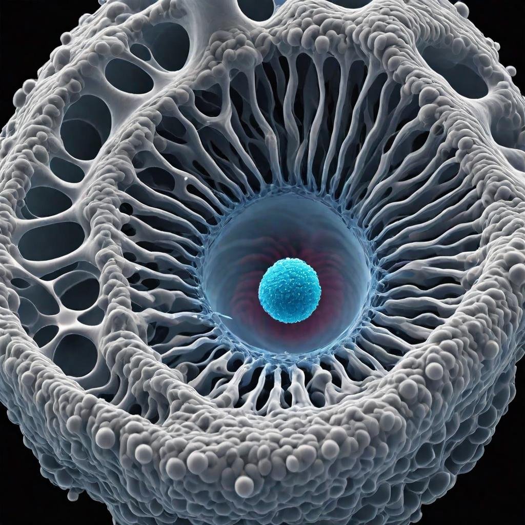
Unlike TEM, SEM creates a detailed three-dimensional image by scanning a concentrated beam of electrons across a sample’s surface. This approach is very effective for investigating surface topographies and compositions. Significant improvements in resolution and depth of field have been made in SEM recently.
Innovations in Electron Microscopy
Significant technological advancements in the field of electron microscopy have increased its areas of application as well as operating in recent years.
Environmental SEM (ESEM)
The development of Environmental SEM, which enables unprepared imaging of specimens in their natural state, is one of the most important developments in electron microscopy. This method is essential for examining hydrated materials and living samples, which would ordinarily degrade in the high vacuum of a conventional SEM.
Cryo-Electron Microscopy (Cryo-EM)
Because it eliminates the need for dyes or stains and allows researchers to study proteins and other biomolecules in their native environments, cryo-EM has revolutionized structural biology. This approach was recently awarded the Nobel Prize in Chemistry for its significant influence on the life sciences. High-resolution biomolecule structures can be obtained using cryo-EM, which helps with drug development and the identification of illness causes.
Focused Ion Beam (FIB)
SEM is often used with focused ion beam technology to allow for accurate manipulation and machining of nanoscale materials. This technology is vital for materials research, electronics manufacturing, and even cultural heritage preservation.
Direct Electron Detectors
These have drastically altered the landscape of electron microscopy by increasing the speed and sensitivity of image acquisition. Direct electron detectors enable real-time imaging and have helped advance dynamic studies that demand great temporal resolution.

Future Prospects and Challenges
The future of electron microscopy seems promising, with continuing research concentrating on enhancing resolution limitations, specimen preparation procedures, and accessibility to these complicated machines. One of the most intriguing developments is the use of artificial intelligence to automate image interpretation, which can dramatically accelerate research and discoveries.
Challenges still exist, particularly in terms of sample preparation and the harm that electron beams can bring to material. However, current research into gentle beam technology and improved sample preparation methods continues to address these concerns.
High-Resolution Techniques
Achieving faster and greater resolution imaging capabilities is one of the most important areas of improvement in electron microscopy. Our capacity to observe atomic structures in previously unheard-of detail has been revolutionized by high-resolution electron microscopy methods like aberration-corrected electron microscopy and High-Resolution Transmission Electron Microscopy (HRTEM). These developments offer insights that direct scientists in the creation of more effective and efficient goods, with significant ramifications for the development of novel materials and nanotechnology.
Automation and Computational Enhancements
Automation has revolutionized electron microscopy, especially with the use of machine learning and sophisticated computational techniques. These advances make strides the precision and speed of information preparing in expansion to computerizing tedious forms. In arrange to discover designs in information that are incomprehensible for individuals to discover, machine learning calculations are being utilized increasingly , driving to modern disclosures and experiences.
This processing capacity also enables electron tomography, a more recent method that creates 3D models of specimens from many 2D images. Thanks to this method, structures at the nanoscale can be understood in three dimensions, which is extremely useful in materials science and biology.
Conclusion
Electron microscopy remains a dynamic area with numerous potential for further investigation and innovation. As we continue to create and improve these powerful tools, our understanding of the microscopic world around us will only grow. Progresses in electron microscopy methods not as it were highlight our require for information, but too give us with made strides implies to handle a few of the foremost challenging logical subjects of our time.
The field of electron microscopy holds incredible guarantee for forming logical and mechanical advancements within the future. The field’s researchers are in for an energizing period as they proceed to investigate the obscure, helped by the continually advancing capabilities of the electron magnifying lens.
FAQs
How is electron microscopy used in medical research?
Understand the role in diagnosing diseases, studying virus structures, and its contributions to biomedical research.
What are some of the latest innovations in electron microscopy?
Get insights into cutting-edge techniques such as cryo-electron microscopy and environmental electron microscopy, and how they are revolutionizing research.
How has electron microscopy impacted healthcare?
It has greatly enhanced the field of virology, pathology, and genetic research by allowing detailed views of the virus structures, cellular components, and other minute particles that are crucial for diagnostics and research.


1 thought on “Electron Microscopy”